If you’re considering ultrasonic cavitation for body contouring — or have already had a session and found yourself typing this question into Google — you’re not alone. “Can ultrasonic cavitation cause cancer?” is one of the most anxiety-driven searches in the aesthetic treatment space, and for good reason: the word “ultrasound” sounds medical, “cavitation” sounds like something happening inside your body, and “cancer” is everyone’s worst-case scenario.
The short answer: no, ultrasonic cavitation does not cause cancer. But the short answer isn’t satisfying when your health is on the line. This article explains why it’s safe — from the physics of ultrasound to the latest clinical evidence — so you can make your decision based on science, not fear.

What Is Ultrasonic Cavitation — and What It Isn’t
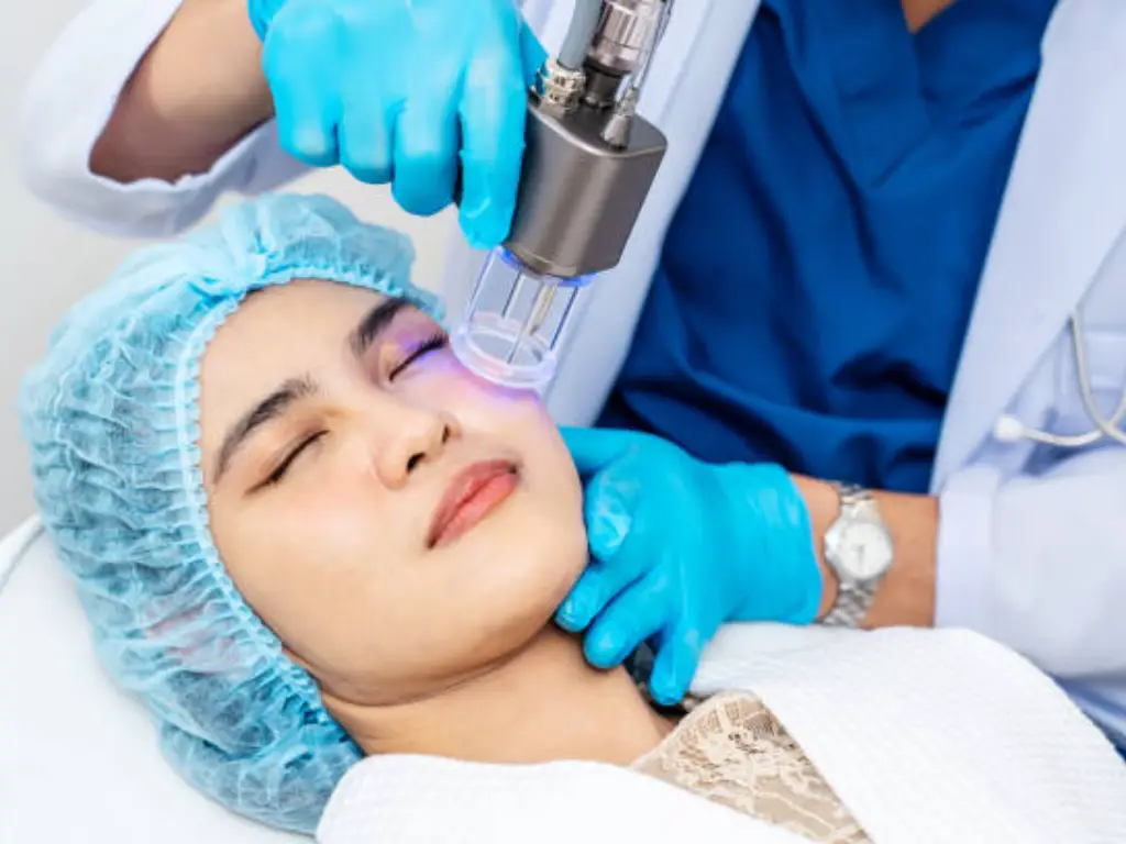
Ultrasonic cavitation is a non-invasive cosmetic procedure that uses low-frequency ultrasound waves (typically around 40 kHz) applied to the skin’s surface to break down fat cells beneath. The ultrasound creates rapid pressure changes in the tissue, forming microscopic bubbles around fat cells. These bubbles expand and collapse — a process called cavitation — mechanically disrupting the fat cell membranes while leaving surrounding tissues like blood vessels, nerves, and muscle intact. Your body then clears the released fat through the lymphatic system and liver over the following days and weeks.
It’s just as important to know what ultrasonic cavitation is not. It is not a weight-loss treatment — it’s a body contouring procedure designed for localized fat pockets in people already close to their goal weight. It is not surgery — no incisions, no anesthesia, no downtime. And critically for our topic, it is not the same thing as the ultrasound your doctor uses for imaging or the high-intensity focused ultrasound (HIFU) used in cancer treatment. Those distinctions matter, and we’ll explore them next.
The typical treatment targets the abdomen, flanks, thighs, and upper arms. Most people need one to three sessions spaced at least 72 hours apart, with visible results appearing over six to twelve weeks as the body gradually processes and eliminates the disrupted fat.
“But I Heard Ultrasound Can Be Dangerous” — Why the Cancer Fear Exists
If ultrasound is safe, why do so many people worry it might cause cancer? The answer lies in a fundamental confusion: not all things called “ultrasound” are the same thing. Think of it like fire — a candle flame, a stovetop burner, and a welding torch all produce heat, but only one of them can melt steel. The energy intensity, how it’s focused, and what it’s used for make all the difference.
Understanding which type of ultrasound you’re dealing with is the first step to separating fact from fear. Below, we break down the three types people most commonly confuse.
Diagnostic Ultrasound — the One Your Doctor Uses
Diagnostic ultrasound is what you encounter during medical imaging — pregnancy scans, abdominal examinations, thyroid checks. It operates at high frequencies (2–18 MHz) but extremely low energy levels, creating real-time images by bouncing sound waves off internal structures.
This is the most studied form of ultrasound in human history. Billions of diagnostic scans have been performed worldwide since the 1950s, and the safety record is extraordinary. A landmark review by Barnett et al. (1997), published in Ultrasound in Medicine & Biology, examined decades of evidence and concluded there is “no indication that medical ultrasound is capable of inducing mutations in mammalian tissue in vivo.” The American Institute of Ultrasound in Medicine (AIUM) reaffirms this position in its current safety guidelines.
Yes, diagnostic ultrasound can produce tiny, transient cavitation effects — microscopic bubbles that form and collapse in tissue. But at diagnostic energy levels, these effects are negligible and tightly controlled. No epidemiological study has ever linked diagnostic ultrasound to increased cancer risk, despite its use on some of the most vulnerable tissue in the human body — developing fetuses. If prenatal ultrasound, applied directly to rapidly dividing fetal cells, shows no cancer signal after decades of global use, the safety precedent is extraordinarily strong.
Therapeutic HIFU — the One That Treats Cancer
At the opposite end of the energy spectrum sits High-Intensity Focused Ultrasound (HIFU). Unlike the broad, low-energy waves of diagnostic scanning, HIFU concentrates ultrasound energy to a millimeter-scale focal point — generating temperatures high enough to thermally destroy targeted tissue.
Here’s the fact that reframes the entire cancer question: HIFU is an FDA-approved cancer treatment. Since 2015, it has been cleared for prostate cancer ablation. It is also approved for uterine fibroids, bone metastases pain relief, and essential tremor. A newer technique called histotripsy uses cavitation itself — the same physical phenomenon used in aesthetic fat cavitation — to mechanically pulverize tumor tissue without heat, and it has entered clinical use for liver cancer.
The logic is straightforward: medical institutions do not use carcinogens to treat cancer. If the physical mechanism of ultrasound were capable of causing malignancies, it could not simultaneously be one of the most precisely controlled tools in the oncology arsenal.
Aesthetic Cavitation — the One You’re Actually Asking About
Aesthetic ultrasonic cavitation sits firmly in the middle of this energy spectrum — and far closer to the safe end. Here’s how the three compare:
| Type | Frequency | Power | Purpose |
|---|---|---|---|
| Diagnostic Ultrasound (imaging) | 2–18 MHz | Milliwatts | Create internal images |
| Aesthetic Cavitation (fat reduction) | ~40 kHz | ~45 W | Disrupt subcutaneous fat cells |
| Therapeutic HIFU (cancer treatment) | 1–7 MHz | Hundreds of watts | Thermally ablate targeted tissue |
Aesthetic cavitation uses frequencies around 40 kHz — much lower than diagnostic or HIFU frequencies — and power output in the tens of watts, not hundreds. It is applied externally to the skin surface, with energy penetrating only 1–3 cm into subcutaneous fat. It cannot reach internal organs, cannot focus energy tightly enough to cause thermal damage beyond the fat layer, and most importantly, its energy is purely mechanical, not ionizing. That last point is the key to understanding why cancer is not a risk, and it deserves its own section.
Ionizing vs. Non-Ionizing: Why Ultrasound Cannot Damage Your DNA
To understand why ultrasonic cavitation does not cause cancer, you need to understand what does cause cancer — and why ultrasound operates in a completely different physical category.
Cancer begins with DNA damage. A cell’s genetic code must be altered — a mutation that disables tumor-suppressor genes or activates oncogenes — for a malignancy to develop. This requires breaking chemical bonds within the DNA molecule, which in turn requires energy input at the molecular level.

How Cancer Actually Starts — the DNA Damage Pathway
The chemical bonds that hold your DNA together have bond energies in the range of 3–5 electron volts (eV). To break these bonds directly, you need particles or photons carrying energy significantly above that threshold. This is exactly what ionizing radiation does: X-rays and gamma rays carry photon energies above 10 eV — enough to eject electrons from atoms, create free radicals, and trigger the chain of molecular damage that can lead to cancer.
The same logic explains why every known carcinogen is dangerous. Ultraviolet light (UV-B, UV-C) has sufficient photon energy to create thymine dimers in DNA — a specific type of bond distortion that leads to mutations. Radon gas emits alpha particles. Tobacco smoke contains dozens of chemical compounds that form DNA adducts. Asbestos fibers cause chronic inflammation that generates mutagenic reactive oxygen species. Every established carcinogen, regardless of its form, converges on the same endpoint: DNA damage.
Now consider ultrasound. If you were to quantum-describe an ultrasound wave — treating it as a stream of phonons rather than a classical pressure wave — the equivalent “photon energy” would be less than 10⁻⁵ eV. That’s six orders of magnitude below the threshold needed to break a DNA bond. Trying to damage DNA with ultrasound energy is like trying to shatter bulletproof glass by throwing ping-pong balls at it. The problem isn’t insufficient force — it’s that the physical mechanism cannot, even in principle, interact with chemical bonds in the way required for mutagenesis.
Ultrasound Energy Is Mechanical, Not Mutagenic
When ultrasound enters tissue, it produces exactly three types of bioeffects — none of which involve the cell nucleus or DNA:
Thermal effect. Ultrasound causes a mild temperature increase in tissue — typically less than 1°C at the intensities used in aesthetic cavitation. To put this in perspective, protein denaturation (the point at which cellular damage from heat begins) requires sustained temperatures above 40°C. A hot shower produces a larger tissue temperature change than an aesthetic ultrasound session.
Cavitation effect. This is the primary mechanism for fat disruption: microscopic bubbles form, oscillate, and collapse, generating localized mechanical forces that tear apart adipocyte (fat cell) membranes. It is a physical, not chemical, process — comparable to shaking a container of liquid until bubbles form and pop. The cell membrane ruptures, but the nucleus is not the target and DNA is not exposed to any mutagenic agent.
Acoustic streaming. The ultrasound wave pushes tissue fluid in the direction of propagation, creating microscopic currents that help transport disrupted fat toward the lymphatic system. This is purely fluid movement — no different in principle from what happens during a massage, just at a microscopic scale.
The AIUM Bioeffects Committee reviewed all three mechanisms in a 2022 safety analysis published in the Journal of Ultrasound in Medicine, examining aesthetic ultrasound specifically. Its conclusion: none of the bioeffects associated with low-intensity ultrasound have a plausible pathway to carcinogenesis. The review specifically noted that the mechanical index (MI) and thermal index (TI) — the two standard safety metrics for ultrasound exposure — remain orders of magnitude below thresholds of concern during aesthetic treatments.
What About Free Radicals? Addressing the One Theoretical Concern
If you dig deep enough into the academic literature, you may find studies from the late 1990s suggesting that ultrasonic cavitation can generate free radicals. This is technically true — under specific laboratory conditions. When cavitation bubbles collapse, the instantaneous temperature inside the bubble can theoretically reach ~5,000 K, creating conditions for sonochemical reactions that split water molecules into hydroxyl radicals.
But here’s what those studies don’t tell you unless you read the full text: these experiments were conducted in cell-free solutions — beakers of water or culture media exposed to high-intensity ultrasound with no biological context. Living tissue is not a beaker. The human body maintains an elaborate antioxidant defense system — superoxide dismutase (SOD), glutathione (GSH), catalase (CAT) — that continuously neutralizes free radicals as a routine metabolic function.
A 2006 study by Olbrisch et al. closed this question decisively. The researchers directly measured free radical production during ultrasound-assisted liposuction — a procedure using higher ultrasound intensities than aesthetic cavitation — in actual human patients. Their finding: “excessive free radicals are not produced during ultrasound-assisted liposuction.” The body’s antioxidant systems cleared any transient radicals before they could accumulate to biologically relevant levels.
In short: the free radical concern is a laboratory artifact, not a clinical reality. The theoretical mechanism doesn’t survive contact with living biology.
What the Evidence Says — Clinical Studies and Real-World Data
Scientific principles are one thing. But you’re entitled to ask: what does the actual data show? If ultrasonic cavitation caused cancer, where is the signal?
Recent Clinical Trials — What 2024–2025 Research Shows
The most recently published clinical evidence comes from a July 2025 study in the Egyptian Journal of Hospital Medicine. Researchers randomized 60 obese adolescent females into three groups: ultrasonic cavitation plus diet, whole-body vibration plus diet, or diet alone. The cavitation protocol used 40 kHz / 45 W — standard aesthetic parameters — twice weekly for six weeks. Results showed statistically significant reductions in body weight, BMI, waist circumference, and abdominal fat thickness in all groups (p < 0.001), with the cavitation group showing superior outcomes. The safety finding: “no significant adverse effects” were reported across the entire treatment period.
Meanwhile, ClinicalTrials.gov lists multiple active trials — including NCT06729203, evaluating ultrasonic cavitation combined with exercise and diet for insulin resistance in centrally obese women — that classify cavitation as a low-risk intervention in their study designs. These trials exclude participants with active cancer or cancer history, not because cavitation is believed to cause cancer, but because researchers apply a universal precautionary principle: any intervention being studied in healthy volunteers excludes individuals whose baseline risk profile complicates safety interpretation.
The Diagnostic Ultrasound Precedent — Billions of Scans, Zero Cancer Signal
The most powerful safety evidence for ultrasound doesn’t come from aesthetic cavitation studies at all. It comes from diagnostic ultrasound. Since the 1960s, obstetric ultrasound has been used on pregnant women — directing ultrasound waves at developing fetuses whose rapidly dividing cells should, in theory, be maximally sensitive to any mutagenic insult. Billions of scans later, no epidemiological study has detected an increased cancer risk in children exposed to prenatal ultrasound.
The World Health Organization has reviewed this evidence and maintains that diagnostic ultrasound, when used appropriately, poses no known cancer risk. If ultrasound waves passing through fetal tissue — the most vulnerable biological target imaginable — show no carcinogenic signal after six decades of global surveillance, the risk from surface-level aesthetic cavitation in adults is, by any rational extrapolation, effectively zero.
When Ultrasound Is Used to Treat Cancer — the Ultimate Irony
We touched on this earlier, but it bears repeating with the evidence: HIFU is an FDA-cleared cancer treatment modality. Prostate cancer, uterine fibroids, bone metastases, and — through histotripsy — liver tumors are all treated using focused ultrasound. The American Cancer Society lists HIFU among recognized focal therapy options for prostate cancer.
To believe that ultrasonic cavitation causes cancer, you would have to believe that the medical community is simultaneously:
- Using ultrasound to cure cancer
- Missing the fact that ultrasound causes cancer despite decades of research specifically looking for bioeffects
That contradiction doesn’t exist in the real world. It only exists in the gap between public understanding and the scientific literature — a gap this article aims to close.
The Real Risks of Ultrasonic Cavitation — What You Should Actually Worry About
No medical or aesthetic procedure is zero-risk, and ultrasonic cavitation is no exception. The real risks are well-documented and almost entirely preventable — but they have nothing to do with cancer.
The common side effects are mild and temporary: redness at the treatment site, slight swelling, occasional bruising, increased thirst as your body processes the released fat, and a buzzing or ringing sensation in the ears during the session itself. These affect a minority of patients and resolve within hours to three days. Some people also report mild nausea that goes away with hydration.
Rare but serious risks are almost universally linked to improper equipment settings or inadequately trained operators. These include:
- Skin burns — when the ultrasound handpiece is held in one position too long or set to excessive intensity
- Seromas — fluid-filled pockets that form under the skin when disrupted fat isn’t evenly absorbed
- Contour irregularities — lumps, bumps, or depressions from uneven fat breakdown, sometimes requiring follow-up treatment
- Nerve irritation — temporary numbness or tingling, rarely permanent
- Liver strain — a theoretical concern if an overwhelming amount of fat is released and processed simultaneously, relevant mainly for those with pre-existing liver conditions
Notice the pattern: every serious complication is operator-dependent, not procedure-inherent. This isn’t a treatment where things randomly go wrong — it’s a treatment where poor execution causes predictable problems. That’s actually good news, because it means choosing the right provider eliminates the vast majority of risk.
Who Should Avoid Ultrasonic Cavitation — Contraindications Explained
Most contraindication lists for cosmetic procedures read like legal disclaimers. Here, we explain the why behind each exclusion, because understanding the medical logic helps you make a genuinely informed decision.
Metabolic clearance issues. Your liver and kidneys process and excrete the fat released by cavitation. If you have liver disease (hepatitis, cirrhosis, fatty liver), kidney disease (stones, transplants, renal impairment), or uncontrolled diabetes, your body may not safely handle the metabolic load. This isn’t a reflection on the procedure’s safety — it’s about your body’s processing capacity. The same logic applies to alcohol: clinics ask you to avoid drinking for 48 hours before and after treatment because your liver is about to get a temporary workload increase.
Blood and circulatory concerns. Cavitation creates microtrauma in fat tissue — that’s the whole point. If you have a coagulation disorder, take blood-thinning medications (warfarin, clopidogrel, even high-dose aspirin), or have active skin infections in the treatment area, the procedure’s mechanical mechanism becomes a risk rather than a benefit. Pacemakers and metal implants are contraindicated because ultrasound vibrations can theoretically interfere with device function, though the risk at aesthetic intensity levels is low.
Special physiological states. Pregnancy and breastfeeding are universal exclusions — not because cavitation is known to be harmful during these states, but because no one has (or should) conduct safety trials on pregnant women. The precautionary principle applies. Similarly, individuals with active cancer or a history of malignancy are typically excluded — again as a precaution, not because cavitation is believed to cause or spread cancer. The theoretical concern is that cavitation-driven lymphatic movement could, in principle, mobilize dormant micrometastases. There is no clinical evidence this actually happens, but the precaution is medically prudent until definitive data exists.
- Liver disease
- Kidney disease
- Uncontrolled diabetes
- Coagulation disorders
- Blood-thinning medications
- Pacemakers / metal implants
- Pregnancy / breastfeeding
- Active cancer history
- General precautionary exclusion
How to Ensure a Safe Ultrasonic Cavitation Treatment
Safety isn’t just about the technology — it’s about who wields it and how. Here are the four lines of defense that separate a safe treatment from a risky one.
| Line of Defense | What to Check | Specific Standards |
|---|---|---|
| Operator Qualifications | Is the technician certified? | Formal training certificate from an accredited aesthetic training provider; ask about course duration and hands-on training hours |
| Equipment Compliance | Is the device cleared for use? | FDA 510(k) clearance or CE marking under Medical Device Directive 93/42/EEC; verify the brand and model online before booking |
| Pre-Treatment Screening | Does the clinic assess contraindications? | A thorough health history questionnaire covering liver/kidney conditions, cancer history, blood clotting, pregnancy status, and implanted devices — not a checkbox form that takes 30 seconds |
| Treatment Protocol | Are the parameters within safe limits? | 40 kHz frequency range (not below 35 kHz), single area treatment ≤ 20 minutes, minimum 72 hours between sessions on the same area |
Beyond the checklist, trust your senses during the treatment. You should feel warmth and possibly a mild tingling sensation, along with the characteristic buzzing sound in your ears. You should not feel sharp pain, burning, or intense discomfort. If you do, speak up immediately and ask the technician to stop. A properly trained operator will check in with you regularly and adjust settings based on your feedback.
For professionals researching ultrasonic cavitation equipment — whether for a clinic, salon, or distribution business — your clients’ safety starts with the quality of the devices you buy. Look for manufacturers that hold ISO 13485 certification and maintain CE and FDA compliance across their product lines. Guangzhou-based Konmison, for example, holds ISO 13485, CE, FDA, RoHS, and FCC certifications and subjects every device to standardized quality checks including 48-hour PCB aging and temperature cycling before shipment. Choosing equipment with a verifiable compliance trail is the most effective step you can take to protect both your clients and your business.
References
- Barnett SB, Rott HD, ter Haar GR, Ziskin MC, Maeda K. “The sensitivity of biological tissue to ultrasound.” Ultrasound in Medicine & Biology. 1997;23(6):805-812.
- American Institute of Ultrasound in Medicine (AIUM) Bioeffects Committee. “Ultrasound for Aesthetic Applications.” Journal of Ultrasound in Medicine. 2022. https://onlinelibrary.wiley.com/doi/10.1002/jum.15856
- Olbrisch RR, et al. “Excessive free radicals are not produced during ultrasound-assisted liposuction.” 2006.
- “Effect of Ultrasound Cavitation versus Whole Body Vibration on Abdominal Fat in Obese Adolescent Females.” Egyptian Journal of Hospital Medicine. July 2025. https://ejhm.journals.ekb.eg/article_453464.html
- “Effect of Ultrasound Cavitation on Insulin Resistance in Patients with Central Obesity in Female.” ClinicalTrials.gov ID: NCT06729203. https://clinicaltrials.gov/study/NCT06729203
- Konmison Quality Assurance. https://www.konmison.com/quality/
- Konmison Cavitation Machine. https://www.konmison.com/cavitation-machine/
- Konmison Homepage. https://www.konmison.com/